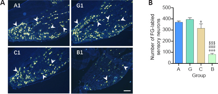
Figure 5.

Effect of the granular hydrogel nerve guidance conduit on the FG-labeled sensory neurons in the DRG of rats with sciatic nerve injury 16 weeks after surgery.
(A) The distribution of the FG-labeled sensory neurons (arrowheads) in DRG. Groups G (G1) and A (A1) showed more labeled neurons than Groups C (C1) and B (B1). Scale bar: 200 µm. (B) The number of FG-labeled sensory neurons on three dorsal root ganglion slices from each animal. Data are shown as the mean ± SD (n = 3). *P < 0.05, ***P < 0.001, vs. Group G; ###P < 0.001, vs. Group A; $$$P < 0.001, vs. Group C (one-way analysis of variance followed by Tukey’s post hoc test). FG: Fluoro-gold; Group A: autologous nerve group; Group B: bulk hydrogel group; Group C: chitosan conduit group; Group G: granular hydrogel group.
