
Figure 6.
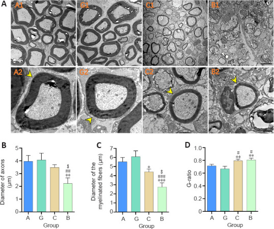
Effect of the granular hydrogel nerve guidance conduit on the morphology analysis of regenerated nerves in rats with sciatic nerve injury 16 weeks after surgery.
(A) The ultrastructure of the regenerated nerves (arrowheads). The diameters of nerve fibers in Groups A (A1 and A2) and G (G1 and G2) were visibly larger and the distribution of myelin sheaths was more compact and uniform than Groups C (C1 and C2) and B (B1 and B2). Scale bars: 5 µm (upper), 1 µm (lower). (B–D) Quantitative results of the diameter of axons (B), the diameter of myelinated fibers (C), and the G-ratio (D) in the distal portion of regenerated nerves. Data are shown as the mean ± SD (n = 3). *P < 0.05, **P < 0.01, ***P < 0.001, vs. Group G; #P < 0.05, ##P < 0.01, ###P < 0.001, vs. Group A; $P < 0.05, vs. Group C (one-way analysis of variance followed by Tukey’s post hoc test). Group A: autologous nerve group; Group B: bulk hydrogel group; Group C: chitosan conduit group; Group G: granular hydrogel group.