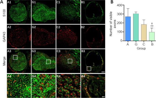
Figure 7.

Effect of the granular hydrogel nerve guidance conduit on the regenerated nerves in rats with sciatic nerve injury 16 weeks after the surgery.
(A) The expression of S100 (green, Alexa Fluor 488) and GAP43 (red, Alexa Fluor 594) in the transverse sections of regenerated nerves. Visibly more myelin sheaths and axons were regenerated in Groups G (G1–G4) and A (A1–A4), followed by Groups C (C1–C4) and B (B1–B4). Scale bars: 100 µm (upper three rows), 20 µm (lowest row). A4, G4, C4, and B4 are the zoomed-in fields of A3, G3, C3, and B3, respectively. (B) Quantification of the number of visible axons. Data are shown as the mean ± SD (n = 3). **P < 0.01, vs. Group G; #P < 0.05, vs. Group A (one-way analysis of variance followed by Tukey’s post hoc test). GAP43: Growth associated protein 43; Group A: autologous nerve group; Group B: bulk hydrogel group; Group C: chitosan conduit group; Group G: granular hydrogel group.
