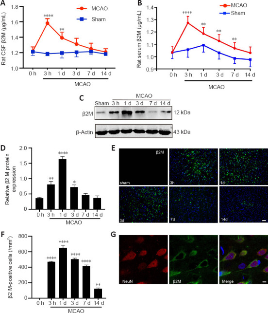
Figure 2.

Cerebral stroke leads to an increase in β2M in rats.
(A, B) β2M in the CSF (A) and serum (B) was detected using ELISA. (C, D) Protein levels of β2M in the ischemic penumbra were quantified. β2M was significantly increased after ischemia. (E, F) Immunofluorescent photomicrographs of β2M (IFKine™ green) and quantitative analysis showed that the number of β2M-positive cells in the ischemic penumbra was also increased in ischemic rats. Data are presented as means ± SEM (n = 4). *P < 0.05, **P < 0.01, ****P < 0.0001, vs. 0-hour group (two-way analysis of variance followed by Bonferroni’s multiple comparisons test for ELISA, one-way analysis of variance followed by Tukey’s honestly significant difference post hoc test for the other data). (G) Immunofluorescent photomicrographs show β2M (IFKine™ green) and NeuN (IFKine™ red) double-labeled cells. Scale bars: 50 μm in E, 2 μm in G. CSF: Cerebral spinal fluid; ELISA: enzyme-linked immunosorbent assay; β2M: β2 microglobulin.
