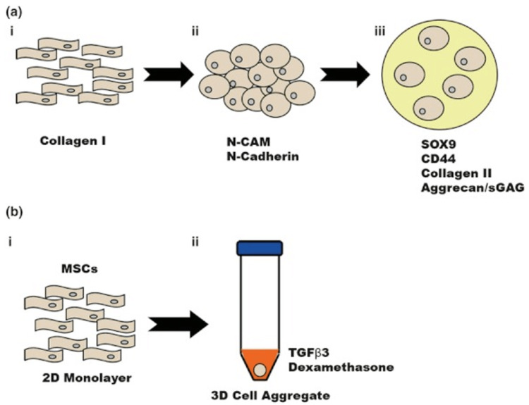
Figure 2. Chondrogenesis of MSCs in vivo and in vitro.
(a) In vivo chondrogenesis. (a.i) Chondrogenesis begins during embryonic development with MSCs expressing collagen I. (a.ii) MSCs migrate and form mesenchymal condensations which activate molecular signalling cascades from plasma membrane receptors, including N-CAM and N-Cadherin. (a.iii) MSCs then proceed to undergo chondrogenic differentiation, depositing cartilage-ECM molecules (such as CD44, collagen II and aggrecan) under the influence of SOX9. (b) In vitro chondrogenesis. (b.i) MSCs are aggregated ex vivo in a 2D-monolayer. (b.ii) MSCs are then added to a 3D-cell aggregate and are grown in this medium. Following this, in vitro differentiation is then the same as in vivo. Adapted from [10] with permission.

