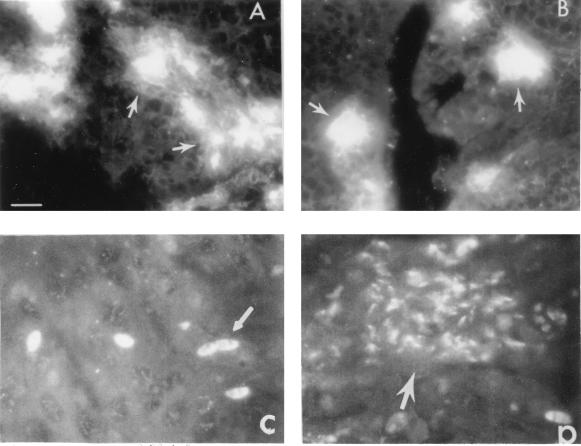
FIG. 2.
Indirect immunofluorescence staining of tissue sections from cecum and cecal tonsils from chickens infected with E. tenella. Developing E. tenella are shown in cecum (A) and cecal tonsil (B) at 72 h postinfection; sporozoites (C) and meronts (D) are shown in cecal tonsil at 24 (C) and 48 (D) h after infection with 105 sporulated oocysts of E. tenella. Each slide contained tissues from three chickens, and three slides were evaluated for the presence of developing parasites. Sporozoites and developing meronts were identified with anti-E. tenella MAb and FITC-conjugated goat anti-mouse IgG and microscopically examined in a Zeiss microscope (bar = 250 μm). The arrows indicate areas with high numbers of intracellular parasites undergoing asexual development (A and B), sporozoites (C), and merozoites (D).

