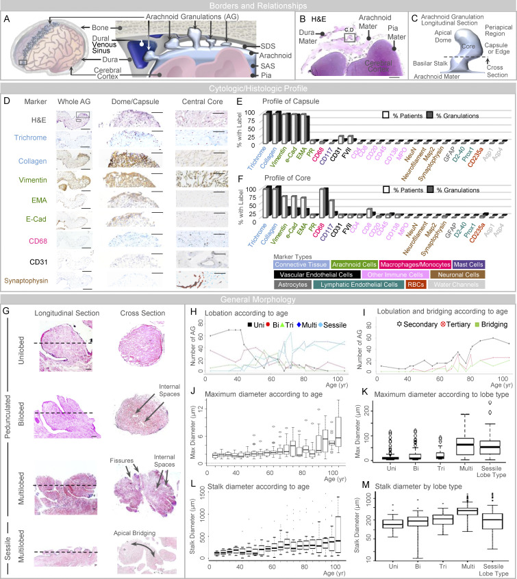
Figure 1.
AG abut dura mater and are heterogeneous in size, form, and composition. (A) A schematic depicts the general arrangement of AG in the superior parasagittal frontal brain region. (B) Similarly, low-power image of an H&E-stained frontal pole section illustrates basic meningeal relationships. (C) Schematic depiction of a single granulation, shown in longitudinal orientation, summarizes its general morphology which includes a capsule or edge, core, basilar stalk or neck, and apical dome region. (D) Intermediate-power images further illustrate labels of an isolated granulation, including dome capsule and central core regions. (E and F) Labeling profiles of each granulation are summarized from all patients at the granulation capsule (or edge) and inner core, respectively, in E and F; cell marker types used for initial screening are shown in the lower panel. (G) As illustrated, AG are pedunculated (or polypoid) and unilobed, bilobed, trilobed, or multilobed; or are sessile and plaque-like in morphology; some granulations exhibit secondary and tertiary lobulations with or without apical bridging. (H and I) The number of morphologies and the number of lobulated and bridged granulations are summarized by age in H and I, respectively. (J and K) Maximum diameter is shown according to age and lobe type in J and K, respectively. (L and M) Stalk diameter is shown according to age and lobe type in L and M, respectively. Scale bars: (B) 500 µm; (D [left]) 200 µm; (D [middle and right]) 50 µm; (G) 20 µm. Bi, bilobed; EMA, epithelial membrane antigen; Multi, multilobed; PR, progesterone receptor; Tri, trilobed; Uni, unilobed. Dotted lines in G represent the approximate orientation of cross-section shown on the right-hand side. Data represent findings in 400 frontal pole granulations from 20 decedents and are from more than two independent experiments.

