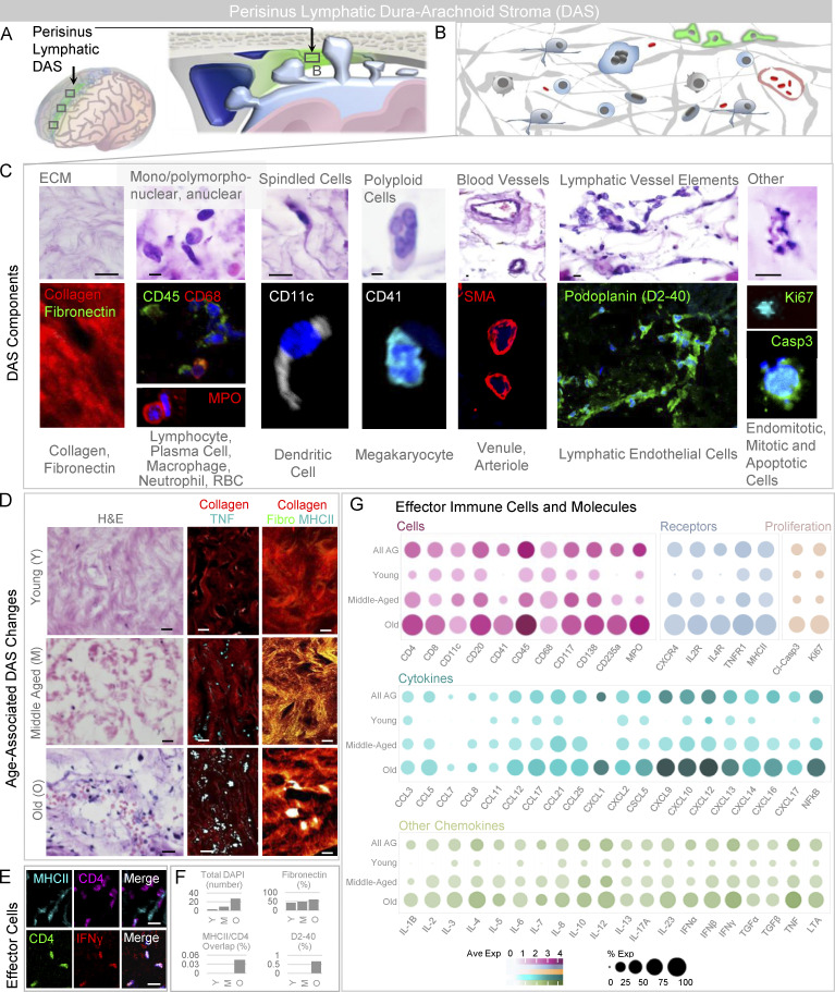
Figure S3.
The perisinus DAS harbors mixed immune cells, immune synapses, and lymphatic endothelial components. (A) The perisinus region houses DAS. (B) This stroma contains extracellular matrix elements, vessels, and heterogeneous cell types. (C) Cropped intermediate-power H&E-labeled and immunofluorescent images of the stromal tissue reveals collagen and fibronectin-rich matrix admixed with immune cells, proliferating and degenerating cells, and blood vascular and lymphatic vascular elements. (D) Intermediate-power images of DAS from young, middle-aged, and old individuals on routine stain (left) and with pan-collagen and TNF label (middle) or pan-collagen, fibronectin, and MHCII labels (right). (E) In addition to trapped TNF-positive cells and cell debris, effector immune cells are observed within DAS of an old decedent. (F) DAS cellularity, percent fibronectin label, percent overlap of MHCII and CD4 labels, and percent D2-40 label are summarized in young (Y), middle-aged (M), and old (O) decedents. (G) Additional cellular, cytokine, chemokine, and molecular label results are summarized according to age, and highlight heterogeneity of immune components in DAS tissue. (C–E) Green or violet/FITC, fibronectin (fibro), CD45, podoplanin, cleaved-caspase 3, Ki67, or CD4; red/CY3, pan-collagen, CD68, myeloperoxidase, SMA, or IFN-γ (γ); white or cyan/CY5, CD11c, CD41, TNF, or MHCII; blue, DAPI. Scale bars: (C and D [right]) 5 µm; (D [left and middle] and E) 10 µm. Data summarize findings in 27 DAS sections from nine decedents and are from >two independent experiments.

