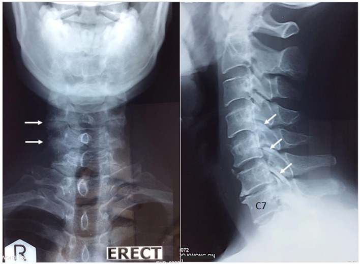
Figure 1.
Anterior and lateral views of the cervical spine. Cervical radiographs show narrowed interspaces from C4/C5 to C7/T1 levels, anterior osteophytic lipping of the cervical vertebrae, degenerative hypertrophy of the facet joints (white arrows), and calcified discs of the C2/C3 and C4/C5 spaces. The radiographic findings are consistent with degenerative spondylosis.

