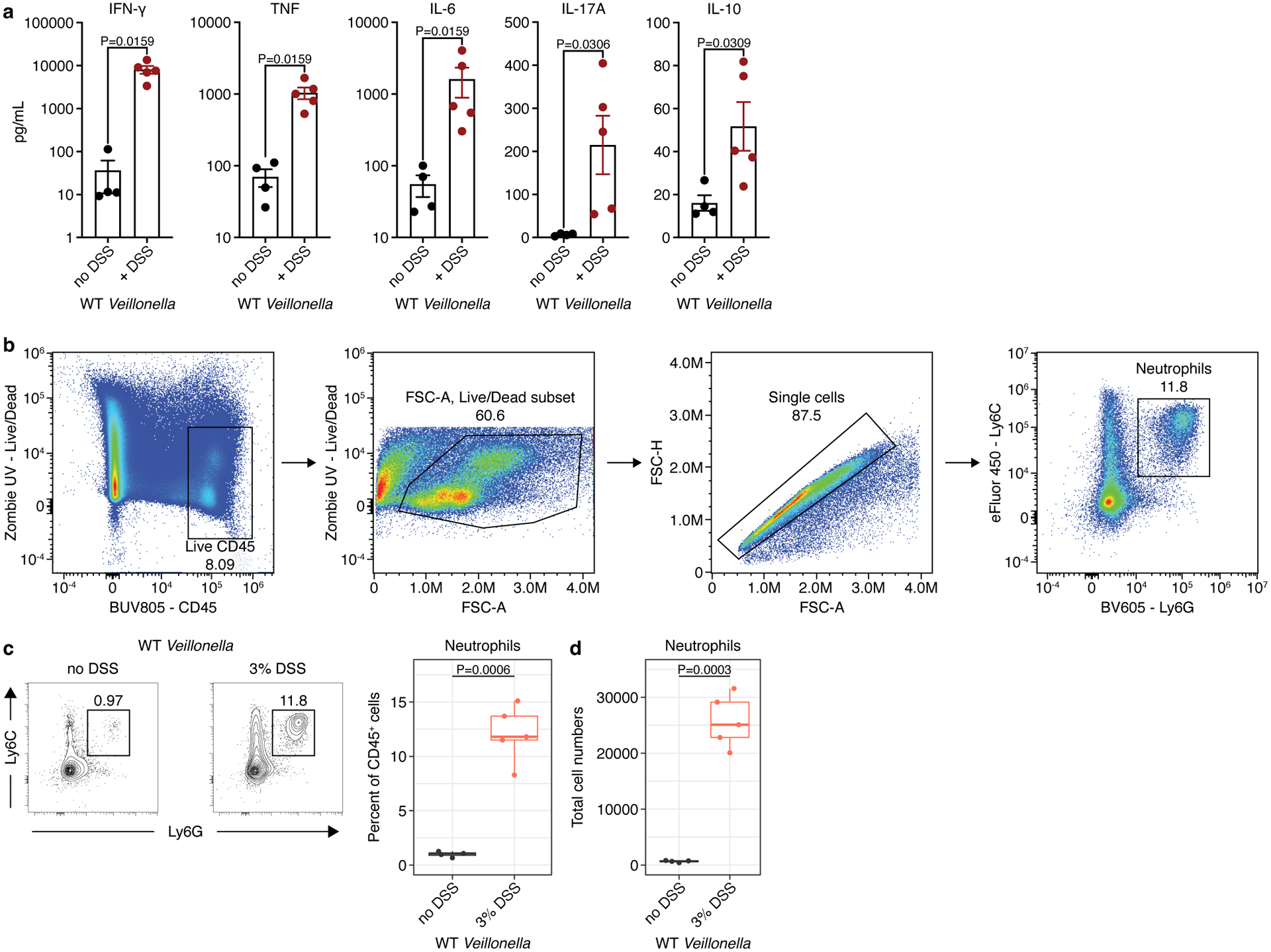
Extended Data Figure 8. Validation of the DSS-treated mouse model.

a) Measurements of IFN-γ, TNF, IL-6, IL-17A, and IL-10 in the mesenteric lymph nodes of WT-colonized ASF mice without (n=4) and with (n=5) DSS treatment. b) Lamina propria cells were isolated from colonic tissue and analyzed via flow cytometry. From left to right: Cells were first gated for positive expression of the pan immune cell marker CD45 while excluding dead cells staining for the viability dye Zombie UV. Next, gating was further cleaned by gating on FSC-A to exclude debris and FSC-A versus FSC-H to exclude doublets. Finally neutrophils were gated based on double positive expression of the two surface protein markers Ly6G and Ly6C. c) Left: representative contour plots showing the proportion of neutrophils based on positive expression of Ly6C and Ly6G on live CD45+ lymphocytes isolated from mouse colonic lamina propria. Right: Frequency of neutrophils among total CD45+ colonic lamina propria lymphocytes. d) Total number of neutrophils isolated from mouse colons based on gating in c. The data in a, c, and d are representative of two independent experiments. For c and d, boxplots display the first and third quartiles with a thick line representing the median; n=4 mice without DSS, and n=5 mice with DSS treatment. A Student’s t-test was used in a, c, and d for statistical analyses.
