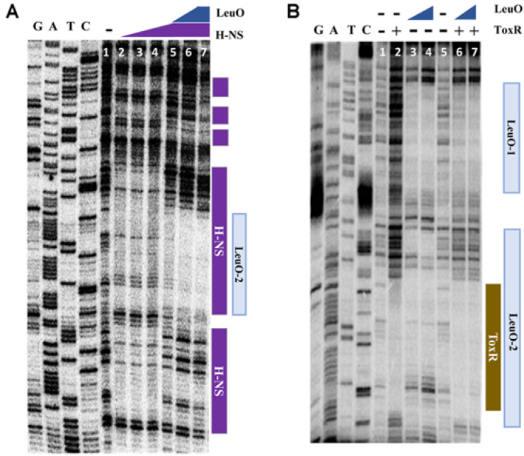
Fig. 5. Comparison of the binding of LeuO, H-NS, and ToxR at various concentrations to the upstream region of leuO.
(A) DNaseI protection assay of the region upstream of leuO using purified LeuO and H-NS; Lane 1, no protein was added; Lanes 2-4, 0.5, 1, and 2 μM of H-NS, respectively, without LeuO; Lanes 5-7, 0.5, 1, and 2 μM of LeuO, respectively, with 2 μM of H-NS. In all lanes, 200 ng of the DNA fragment with 3’- labeled leuO promoter region was added. (B) DNaseI protection assay of the region upstream of leuO using purified LeuO and ToxR; Lanes 1 and 5, no protein was added; Lanes 2, 3 μM of ToxR; Lanes 3-4, 2 μM and 5 μM of LeuO, respectively, without ToxR; Lanes 6-7, 2 μM and 5 μM of LeuO, respectively, with 3 μM of ToxR were incubated. In all lanes, the DNA fragment with 200 ng of 3’- labeled leuO promoter region was added.

