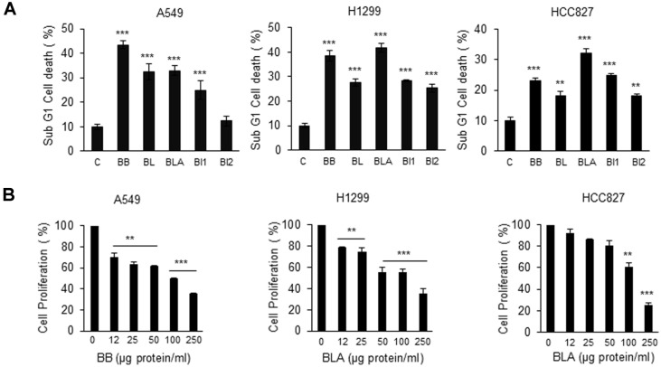
Fig. 1. The total aqueous extract of Bifidobacterium species induced cell death and inhibited proliferation of NSCLC cells.
(A) A549, H1299 and HCC827 (6 × 10⁵) strains were seeded in a 6-well plate. Bifidobacterium bifidum (BB), Bifidobacterium longum (BL), Bifidobacterium lactis (BLA), Bifidobacterium infantis 1 (BI1), and Bifidobacterium infantis 2 (BI2) were applied as aqueous extracts (150 μg protein/ml) and incubated with cells for 24 h. PBS served as a vehicle. The WST- 8 assay was performed to assess cell death. Values are expressed as the mean ± SD (**p < 0.01 and ***p < 0.001, as compared with the PBS treated Control). (B) A549, H1299 and HCC827 cells (6 × 10⁵) were seeded in a 6-well plate. BB or BLA aqueous extracts were added at 0, 12, 25, 50, 100, and 250 μg protein/ml on the following day, for 12 h incubation. WST-8 assay was performed to test cell proliferation. Values are expressed as the mean ± SD (**p < 0.01 and ***p < 0.001 as compared with the PBS treated Control).

