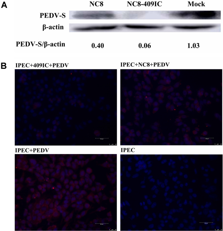
Fig. 7. Recombinant L. plantarum NC8-409IC and NC8-409p’I suppress PEDV infection in IPEC-J2 cells (at the protein level).
IPEC-J2 cells infected with PEDV at 1000× TCID50 for 36 h were assessed by western blot for the PEDV S protein following incubation with NC8-409IC for 2 h (A). IPEC-J2 cells were stimulated with NC8-409IC (at a ratio of recombinant protein to IPEC-J2 cells of 1:20) for 2 h before infection with PEDV for 2 h and were then incubated in maintenance medium for 36 h and fixed with 10% paraformaldehyde. Virus infection was assessed via an IFA for the PEDV S protein. Samples were subjected to staining for PEDV antigen (red). Nuclei were stained with DAPI (blue). The scale bars represent 50 μm (B).

