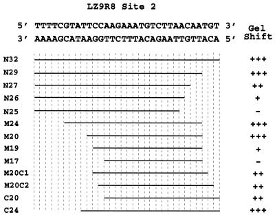
FIG. 12.
Summary of rEUO binding to deletion fragments of LZ9R8 site 2. The 32-bp rEUO binding site, as determined by DNase I protection studies, is shown at the top. Gel mobility shift analysis was carried out with synthetic double-stranded oligonucleotides, deleted from either or both ends of the site, and 1, 10, or 50 ng of rEUO, as described in Materials and Methods. The lengths of fragments are represented by horizontal lines. The gel shift phenotype was scored as follows: +++, shift equal to that of the wild type at all three rEUO concentrations; ++, decreased shift at 1 ng of rEUO, wild-type shift at higher concentrations; +, decreased shift at 1 and 10 ng of rEUO, wild-type shift at 50 ng; −, no major band shift at any of three rEUO concentrations.

