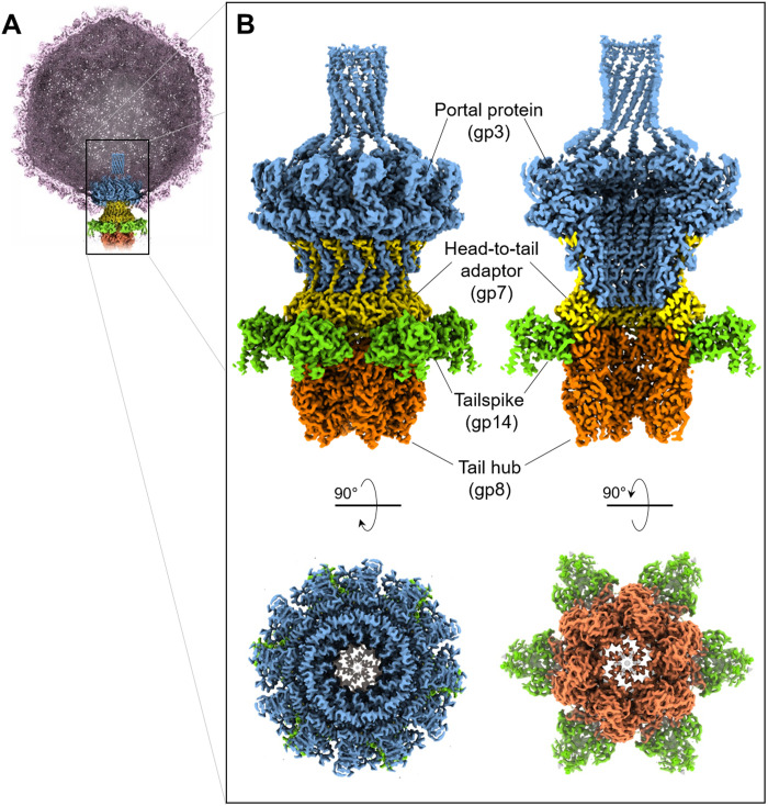
Fig. 1. Localized reconstruction of phage Sf6 tail machine.
(A) A section through the Sf6 virion was visualized at ~1.6σ with the coat protein colored in light magenta. The C6-symmetrized localized reconstruction was overlaid to the unique vertex and is magnified in (B). Tail factors visible at high contour are the portal protein (blue), head-to-tail adaptor (yellow), tailspike N termini (green), and tail hub (orange).

