
Figure 2.
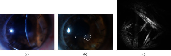
Slit-lamp photograph of the right eye at initial presentation. (a) A slit-beam photograph centered over the new crystalline pattern of branching thin structures in the anterior stroma. (b) A wide-beam photograph demonstrating the branch-like structures in the anterior stroma of the central visual axis (arrowhead), and nasally, a preexisting triangular anterior stromal scar (dashed outline). (c) Confocal microscopy revealed mid-stromal (195 μm deep) highly reflective needle-like structures consistent with crystalline keratopathy, but not specific for an individual pathogen.