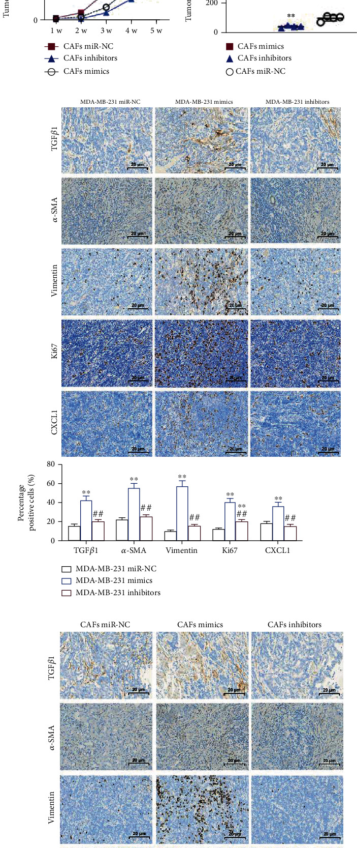
Figure 6.

Exosomal miR-425-5p stimulated tumor growth in vivo. (a) Tumors in mice transplanted with miR-NC transfected MDA-MB-231 cells and miR-425-5p mimics/or inhibitors transfected MDA-MB-231 cells. (b) Growth curve of tumors in different groups. (c) Tumor weight in different groups. (d) Tumors in mice transplanted with miR-NC transfected CAFs and miR-425-5p mimics/or inhibitors transfected CAFs. (e) Growth curve of tumors in different groups. (f) Tumor weight in different groups. (g, h) IHC staining showed the expression of TGFβ1, α-SMA, vimentin, Ki67, and CXCL1 in different groups. Experimental data presented as means ± standard deviation. Experiment was repeated 3 times. ∗∗P < 0.01 compared with the MDA-MB-231 miR-NC/CAFs miR-NC group. ##P < 0.01 compared with the MDA-MB-231 mimics/CAF mimics group.
