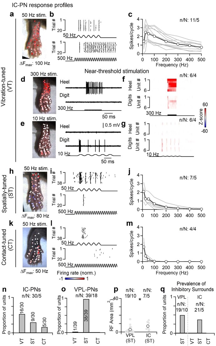
Extended Data Fig. 2. IC projection neuron subtypes.
The DCN is composed of several types of projection neurons and local interneurons. VPL-PNs are the most abundant, with an estimated proportion of VPL-PN:IC-PN:Vgat-IN of 2:1:1 based on retrograde tracing and genetic labeling in mice13. The proportion of VPL-PNs may be larger12. Units that could be antidromically activated from the IC fell into three functional types with distinct properties. Vibration-tuned (VT) units have very large receptive fields that include almost the entire hindlimb, they can entrain their firing to very high frequencies (>300 Hz and often up to 500 Hz), and do not have inhibitory surrounds. The broad receptive field and entrainment to high frequencies suggest that they receive strong input from Pacinian corpuscles. Spatially-tuned (ST) units have small receptive fields, typically consisting of 1-2 digits or pads, or a small portion of the heel. They often have inhibitory surrounds, and can entrain their firing to modest vibration frequencies (up to 100–200 Hz). These units cannot entrain their firing to vibration frequencies > 300 Hz, even at high forces. Contact-tuned (CT) units have receptive field sizes similar to spatially-tuned units, but are poorly activated by vibration. They are very sensitive and almost exclusively responsive to the initial contact of a stimulus. a, Example receptive field of a vibration-tuned IC-PN. Color scale of normalized firing rate shown at bottom. b, Example rasters of a vibration-tuned IC-PN unit in response to 50 (top) or 500 Hz (bottom) vibration. c, Responsiveness of vibration-tuned IC-PN units to different vibration frequencies (10–20 mN). Data is shown for individual units (gray) and average across units (black). d–g, Vibration-tuned units had different sensitivities to different frequencies of vibration. When reducing the force of vibrations to near-threshold (<10 mN), 300 Hz vibrations evoked more robust responses in the heel, whereas 10 Hz vibrations typically only evoked responses when stimulating the digits. d, Receptive field map of near-threshold 300 Hz vibration for an identified vibration-tuned IC-PN (left). Example trials from the heel (top right) and digits (top left) are shown. Color scale of normalized firing rate shown at bottom. e, Same as in d, but for 10 Hz near-threshold vibration. Color scale of normalized firing rate shown at bottom. f, Average histograms for individual units of near-threshold 300 Hz vibration delivered to the heel (top) or digits (bottom). Color scale of Z-scores shown at right. g, Average histograms for individual units of near-threshold 10 Hz vibration delivered to the heel (top) or digits (bottom). Color scale of Z-scores shown at right. h–j, Same as a-c, but for spatially-tuned units. k–m, Same as a–c, but for contact-tuned units. n, Distribution of functional types for units antidromically activated from the IC. o, Functional types were generalized to classify VPL-PNs. Units antidromically activated from the VPL were almost all spatially-tuned. Their receptive fields were small and did not phase-lock to high frequency vibration (>300 Hz), even when increasing the amplitude. These units also typically did not elevate their firing in response to high-frequency vibration. 1/39 units were found to be vibration-tuned. p, Receptive field area for spatially-tuned units antidromically activated from the IC or VPL. Distributions are significantly different (p = 0.031, Kolmogorov-Smirnov test). Individual units (gray) and mean ± s.e.m. (black). q, Proportion of units with inhibitory surrounds for each functional type. All data shown as n units in N animals (n/N).

