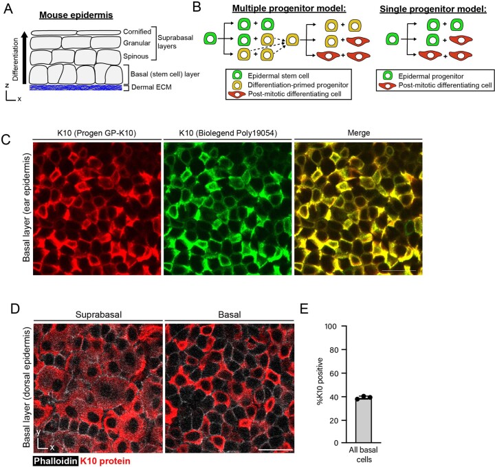
Extended Data Fig. 1. Characterization of Keratin 10 protein in basal cells.
(A) Cartoon schematic of epidermal structure. Stem cells reside in an underlying basal layer (directly above dermal extracellular matrix, shown in blue), and differentiate upwards to contribute to the outer barrier layers of the skin. (B) Current models of epidermal homeostasis propose that the basal layer is composed of either multiple distinct stem/progenitor cells (one common model proposes slow cycling stem cells that give rise to a self-sustaining population of differentiation-primed progenitors; left cartoon) or a single type of epidermal progenitor (right cartoon). (C) Comparison of Keratin 10 whole mount immunostaining in ear epidermis using two independent antibodies (Progen GP-K10 in red; Biolegend Poly19054 in green). Scale bar=10 µm. (D) Representative whole mount staining of Keratin 10 (red) in suprabasal and basal cells from dorsal epidermis. Cell boundaries are visualized with phalloidin (white). Scale bar = 25 µm. (E) Quantification of percent dorsal basal cells that express Keratin 10 protein. Graph represents average from one independent immunostaining experiment using n=3 mice. Error bars are mean ±S.D.

