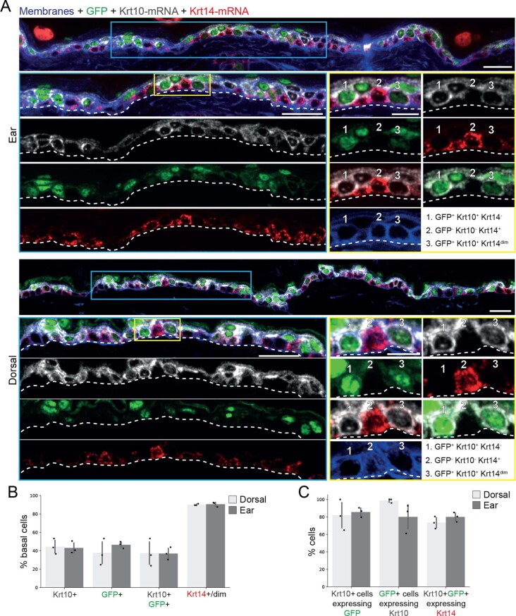
Extended Data Fig. 2. Characterization of Keratin 10 transcripts in basal cells.
(A) Tissue staining for GFP protein (K10rtTA; pTRE-H2BGFP) together with smFISH of Krt10 and Krt14 mRNA in ear and dorsal skin from the same genetic model used for intra vital imaging. Cell membranes were stained with WGA. Numbered cells in the zoom-ins show examples of cells with (1) Krt10-GFP protein and Krt10 mRNA co-expression; (2) Krt14 mRNA expression without Krt10-GFP nor Krt10 mRNA expression; (3) Krt10-GFP protein, Krt10 mRNA and Krt14 mRNA (dim) co-expression. Scale bar = 25 μm (for overviews) or 10 μm (for zoom-ins). (B-C) Quantification of GFP, Krt10 and Krt14 expressing basal cells among all basal cells (B) and their respective co-expression within the same cells (C). n = 3 mice, error bars are mean ±S.D.

