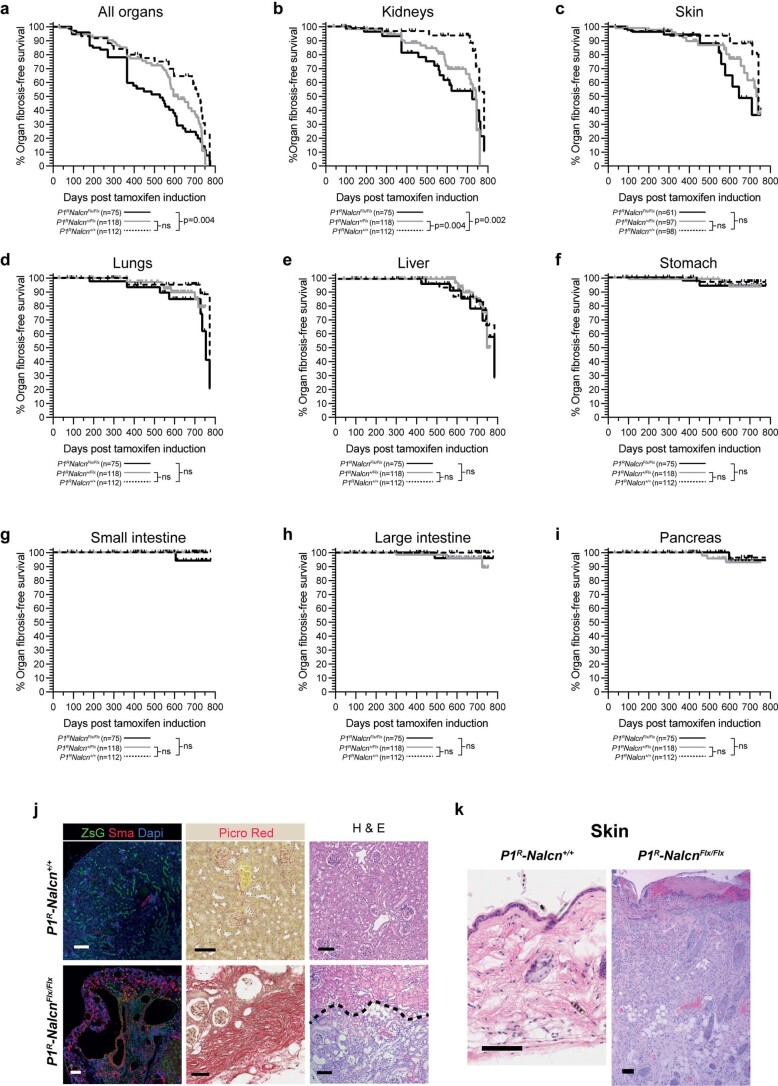
Extended Data Fig. 7. Organ fibrosis following conditional deletion of Nalcn at P3 in P1R mice.
Fibrosis-free survival for all organs (a) or the indicated organs (b-i). P value reports the log-rank statistic (Mantel-Cox). The numbers of animals of each genotype are shown. p-values for each graph comparing P1RNalcn+/+ and P1RNalcn+/Flx and P1RNalcn+/+ and P1RNalcnFlx/Flx, respectively: All organs (0.0664, 0.0035), Kidneys (0.0037, 0.0022), Skin (0.1195, 0.0569), Lungs(0.3791, 0.1000), Liver(0.8846, 0.7250), Stomach(0.4938, 0.4225), Small intestine(>0.9999, 0.2348), Large intestine(0.1312, 0.2655), Pancreas(0.4764, 0.7571). (j) Photomicrographs of haematoxlin and eosin (H & E) and Picro-Sirus Red stain and co-immunofluorescence of ZsGreen, alpha-smooth muscle actin (ASMA) and Dapi in kidney from P1RNalcn+/+ and P1RNalcnFlx/Flx mice aged >400 days. Note in P1RNalcnFlx/Flx kidney: extensive fibrosis below the hashed line (H & E); marked Picro-Sirius Red staining indicating extensive fibrosis; ZsGreen recombination, gross distortion of normal kidney architecture and extensive alpha-SMA expression. (k) Photomicrographs of H & E stained skin from P1RNalcn+/+ and P1RNalcnFlx/Flx mice aged >400 days. Note in P1RNalcnFlx/Flx skin: ulceration and thickening of cornified layer, marked thickening of squamous cell layer and fibrosis of dermal layer. (scale 100um).

