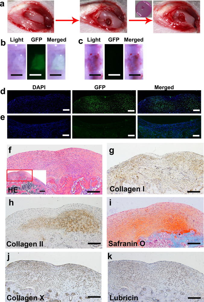
Fig. 6. ECCs can repair knee defects and form layered cartilage structures.
a Schematic representation showing that a full-thickness rat cartilage defect was created. ECCs were seeded in collagen sponges, cultured in vitro for 4 weeks, and then surgically transplanted into cartilage defects. b, d Knees treated with GFP-expressing ECCs loaded on a collagen sponge were harvested 4 weeks after transplantation. GFP expression was detected in the ECC group. b Scale bar, 1 mm. d Scale bar, 50 μm. c, e No GFP expression was observed in the control group transplanted with only the collagen sponge. c Scale bar, 1 mm. e Scale bar, 50 μm. f HE staining showing that the repaired tissue was cartilage-like. (Inset) HE staining at low magnification. Scale bar, 100 μm. (Inset) Scale bar, 200 μm. g Immunohistochemical staining revealing that collagen I was present throughout the cartilaginous cell layers. h Immunohistochemical staining showing that collagen II was predominant in the deep layer. i Safranin O is more significantly positive than the deeper layer. j, k Immunohistochemical staining showing that collagen X and lubricin are expressed in the regenerated cartilage tissue. g–k Scale bar, 100 μm.

