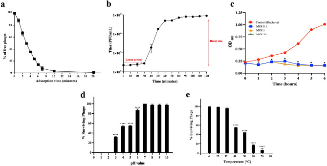
Figure 4.
In vitro characterization of the phage vB_PaeS_VL1. (a) Kinetic adsorption of the phage to the host strain (P. aeruginosa ATCC27853) at an MOI of 0.01. (b) One-step growth curve of phage VL1 at a MOI of 0.001. (c) Bacterial killing activity of phage VL1 against host cells at different MOI (0.1, 1, and 10) in TSB medium. (d) Phage VL1 stability under different pH conditions. (e) Stability of Phage VL1 under different temperature conditions. Data are presented as mean ± SD of three independent experiments. The asterisks indicate significant differences (*P < 0.05 and ****P < 0.0001, one-way ANOVA followed by Dunnett's post hoc test) between the experimental and control groups.

