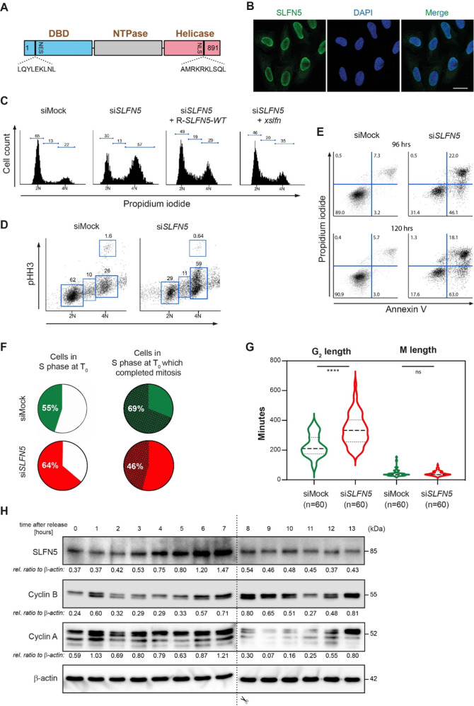
Fig. 1. SLFN5 loss-of-function results in G2/M arrest.
A Structure of SLFN5. SLFN5 consists of three hitherto uncharacterized domains: a N-terminal DNA binding domain (DBD), a C-terminal DNA/RNA helicase domain and a nucleoside triphosphate hydrolase domain (NTPase). A canonical nuclear localization signal (NLS) and a nuclear export signal (NES) are present at the C- and N-terminus, respectively. B Immunostaining of U2OS cells with #111/1 SLFN5 monoclonal antibody reveals nuclear localization of SLFN5. DNA is stained with Hoechst. Scale bar represents 20 µm. C, D Downregulation of SLFN5 in U2OS cells by RNAi for 48 h leads to G2/M arrest. G2/M arrest after siRNA-mediated SLFN5 knockdown can be partially rescued by a siRNA-resistant (R-)SLFN5 construct and its Xenopus laevis ortholog xslfn. Cells were harvested 48 h after transfection with siMock or siSLFN5. Rescue experiments were performed by concomitant transfection of siSLFN5 and R-SLFN5 or xslfn. Cell cycle analysis was performed by combined propidium iodide (PI)/phosho-histone H3 (pHH3) FACS staining. The pictures show a representative experiment from three independent experiments. E Downregulation of SLFN5 in U2OS cells by RNAi leads to apoptosis. Cells were harvested 96 and 120 h after transfection with siMock or siSLFN5. The fraction of apoptotic cells was determined after Annexin V staining by FACS analysis. The pictures show a representative experiment from three independent experiments. F, G Cell cycle progression of SLFN5 depleted cells assessed by time-lapse microscopy over 12 h. Pie charts indicate percentages of cells in S phase at T0 (start of filming) (F, left) and cells in S phase at T0 which successfully divided (F, right), respectively. Violin plots statistically illustrate length of both G2 and M phase of SLFN5-depleted cells G. Green and red colours identify siMock-treated and siSLFN5-treated cells, respectively. The graphs statistically illustrate the results from three independent experiments. H Western blotting of SLFN5 expression during cell cycle in U2OS cells. U2OS cells were arrested at G1/S phase by double-thymidine treatment and released from arrest by thymidine washout. Samples were collected for Western blotting in 60 min intervals, until cells reached the mitotic state (12–13 h). Cyclin B and Cyclin A were used as markers for G2/M and interphase, respectively. β-actin was used as loading control. The Western blot shows a representative experiment from three independent experiments.

