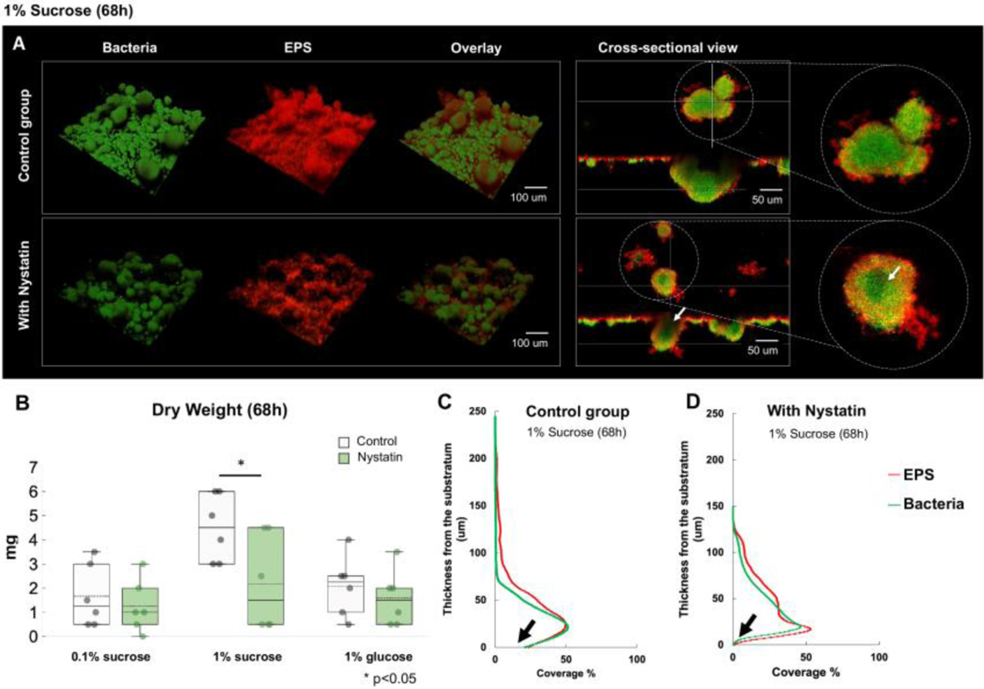
Fig. 3. Morphogenesis and 3D architecture of S. mutans and C. albicans duo-species biofilms treated by Nystatin.

(A) Morphogenesis and 3D architecture of 68h Nystatin-treated biofilms were visualized. Altered biofilm structures were seen, with smaller and hollow microcolonies. White arrow indicates inner aspect of microcolony. (B) Significant reduction of biofilm dry weight following Nystatin treatment was seen in 1% sucrose condition, comparing to the control group, (p=0.04, t-test). (C-D) Layer distribution of the 68h biofilms formed in 1% sucrose showed that the Nystatin-treated biofilms had thinner layers and with less biomass at the substrate layer (marked with black arrows).
