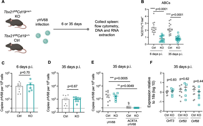
Figure 4.
ABCs are not required for controlling lytic infection and achieving long-term latency. Tbx21fl/flCd19+/+ (Ctrl, filled grey circles) and Tbx21fl/flCd19cre/+ (KO, open blue squares) mice were infected i.p. with γHV68 or ACRTA-γHV68. 6 or 35 days p.i., spleens collected and processed for RNA extraction, DNA extraction, and flow cytometry. (A) Experimental scheme for data shown in panels (B–D,F). (B) Proportion of ABCs (CD11c+T-bet+) of previously activated B cells (CD19+IgD−) in the spleen in Ctrl and KO mice 6 and 35 days post-γHV68 infection. (C,D) Splenic γHV68 viral load (copies Orf50 per million cells) in Ctrl (filled circles) and KO (open squares) mice as determined by qPCR day 6 and 35 p.i., with limit of detection indicated by dotted line. (E) Splenic γHV68 viral load (copies Orf50 per million cells) in Ctrl and KO mice infected with γHV68 or ACRTA-γHV68 35 days p.i. (F) Relative expression in the spleen of Orf73, Orf50, and Orf68 in Ctrl and KO mice at day 35 p.i. Each data point represents an individual mouse. Both male and female mice were included in the experiments. Data presented as mean ± SEM, analyzed by Mann–Whitney test (B–D,F) or Kruskal–Wallis H test (E).

