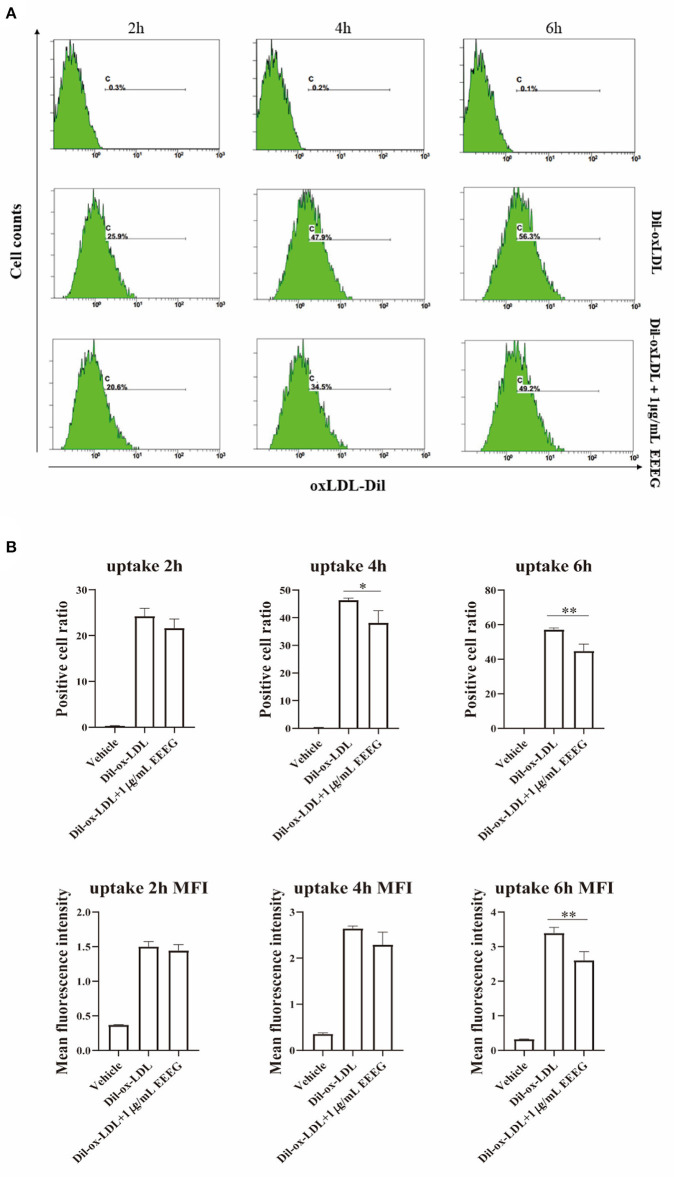
Figure 3.
EEEG inhibits the uptake of oxidized low-density lipoprotein by macrophages. (A) Representative histogram plots of Dil-ox-LDL binding by macrophages. Cells were incubated for 2, 4, and 6 h with Dil-ox-LDL in the presence or absence of EEEG, followed by flow cytometry. (B) Quantification of the percentage of positive cells and mean fluorescence intensity values. DiI-ox-LDL-fluorescence is shown on the Y-axis and macrophage numbers on the X-axis. Data are expressed as mean ± s.e.m., Statistical analysis was based on Graphpad Prism 9.0.2 software and a value of P < 0.05 was considered statistically significant. One-way ANOVA with Student Neuman-Keuls post-hoc test was performed to compare the data between multiple groups, *p < 0.05, **p < 0.01 vs. ox-LDL.

