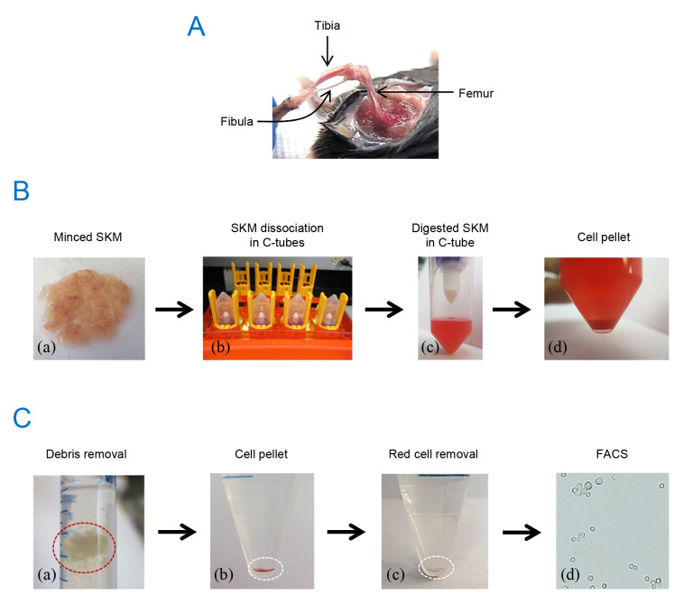
Figure 1. Outline of macrophage isolation from mouse skeletal muscle.
(A) Hind limb after muscle harvest. (B) Minced muscle from mouse hind limbs (a), cell dissociator (b), digested muscle in a C-tube (c), and cell pellet after centrifugation (d). (C) Debris removed by the debris removal solution (a), cell pellet after debris removal (b), cell pellet after red cell removal (c), and cells after sorting (d, 20× brightfield image).

