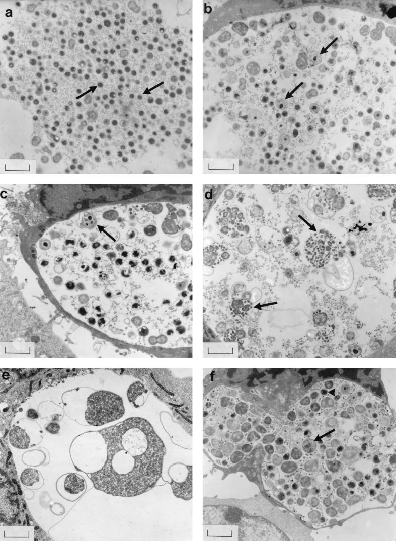
FIG. 1.
Strain 434 inclusions in McCoy cells supplied with medium containing reduced AA concentrations (a to e) or AAs at the concentrations found in blood plasma (f), as viewed by electron microscopy at 40 h p.i. No CH was present. (a) Inclusions in CMEM (100% AAs) contained mainly normal EBs (arrows). (b) AA reduction to 75% of the concentration in CMEM was associated with swollen intermediate forms (arrows). (c to e) Organisms became larger and more distorted as the medium AA supply decreased to 40, 25, and 0%, respectively; individual large forms often contained small particles (particularly noticeable in panel d) (arrows). (f) Many abnormally large chlamydiae (arrow), as well as some normal EBs (arrowhead), were also present in inclusions supplied with blood plasma AA levels Bars = 1.33 μm.

