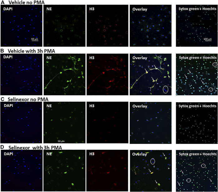
FIGURE 2.
Inhibitory effect Selinexor on NETs formation, as seen in microscopy. Immunofluorescent (IF) staining demonstrating a significant inhibition of NETs formation following incubation of neutrophils with 50 nM Selinexor for 2 h vs. vehicle. Cells were activated with 100 nM PMA for 3 h and compared to non-activated cells. (A) Neutrophils were incubated with vehicle and were not activated. No NETs release was seen (B) Neutrophils were incubated with vehicle and activated with PMA. Significant NETs release was seen. (C) Neutrophils were incubated with Selinexor and were not activated. No NETs release was seen. (D) Neutrophils were incubated with Selinexor and activated with PMA. Minor NETs release was seen. Columns, from left to right: DAPI nuclear staining (blue), Neutrophil elastase (green), Histone 3 (red) and the Overlay of all three images. On the right columns Sytox green and Hoechst 33342 double staining. Representative NETs-forming neutrophils are indicated with white arrows, while neutrophils not forming NETs are signed with white circles.

