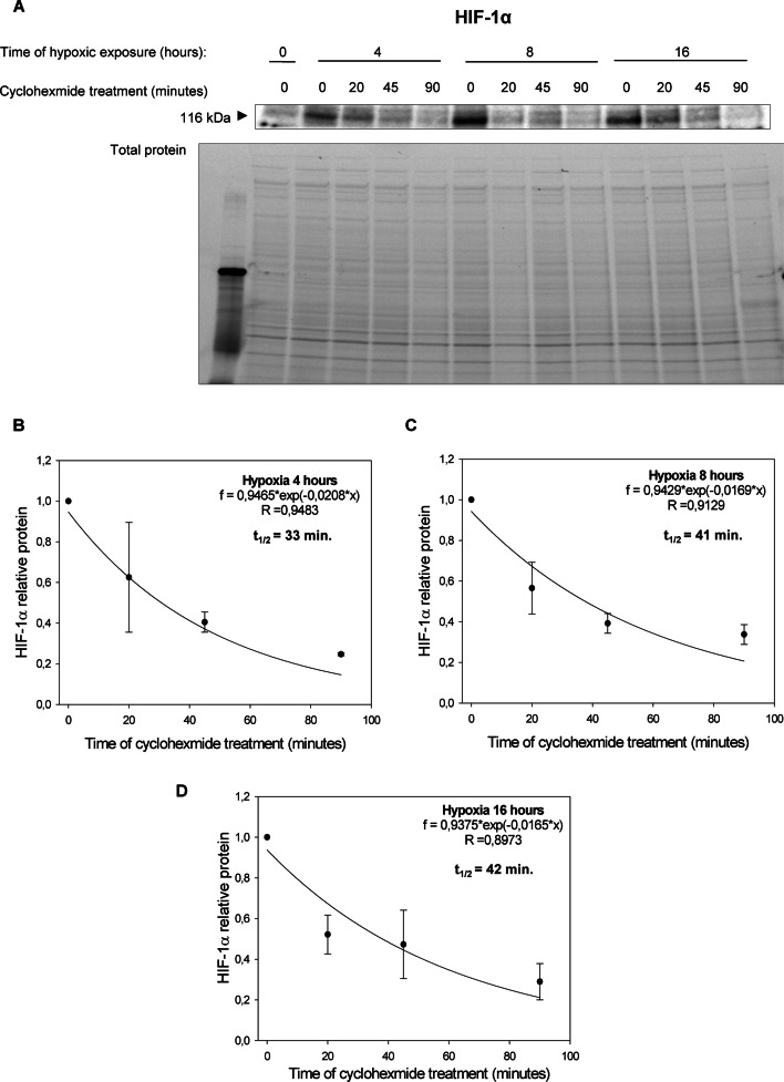
Fig. 2.
Hypoxia effect on HIF-1α protein half-live. A HIF-1α half-live measurements were taken in HUVECs exposed to hypoxia for 4, 8, and 16 h (cells cultured in normoxia were the negative control). Cycloheximide was added to stop translation, after which protein lysates were collected, and HIF-1α levels at each time point were measured by western blot and normalized to total protein levels. Values for each time point were calculated from three individual samples generated in at least three independent experiments. The mathematical representation of HIF-1α protein levels for 4 h (B), 8 h (C) and 16 (D) hypoxia in HUVECs. The time points indicating relative HIF-1α levels were plotted as differences from HIF-1α levels at the initial time point (t = 0). The protein half-lives were calculated from the exponential decay using the trend line equation C/C0 = e–kdt (where C and C0 are protein amounts at time t and at the t0, respectively, and kd is the protein decay constant). The error bars represent SD. * P < 0.05 was considered significant

