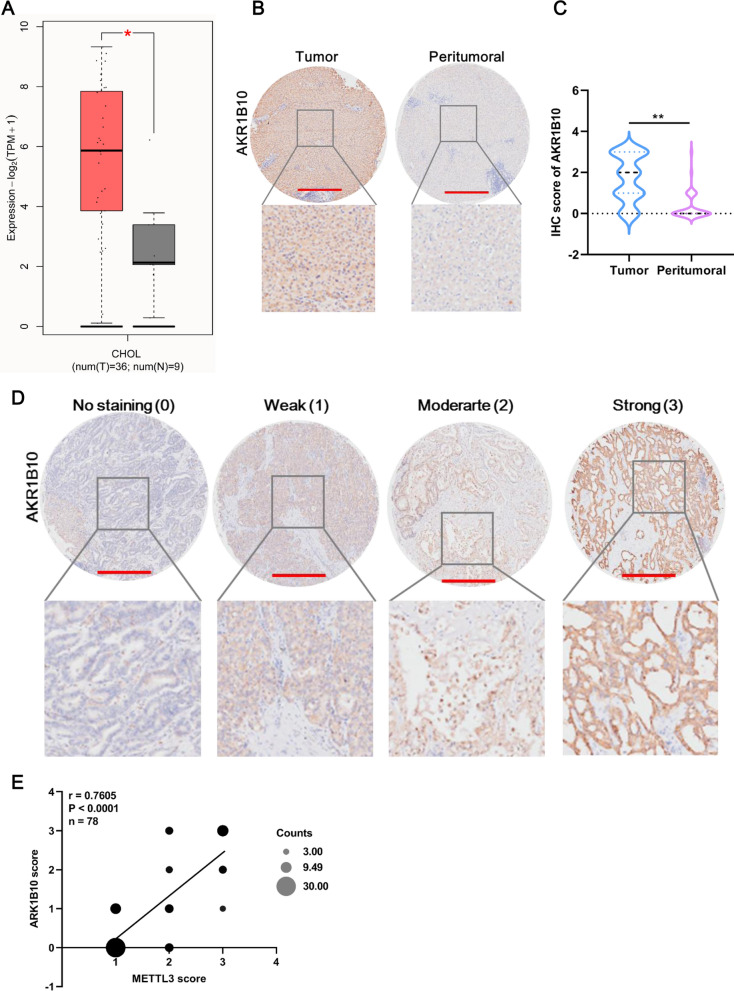
Fig. 6.
AKR1B10 is highly expressed in CCA tissues. A AKR1B10 expression in the GEPIA2 dataset. The red box indicates tumor tissue, and the gray box indicates normal tissue. B Representative image of AKR1B10 expression in tumor and peritumoral tissue in the CCA tissue microarray. Compared with that in peritumoral tissues, AKR1B10 was overexpressed in CCA tissues, as revealed by IHC staining for AKR1B10 in a CCA tissue microarray (n = 60). Scale bar: 500 μm. C The AKR1B10 score in CCA tissues was significantly higher than that in peritumoral tissues. The sample sizes were 60 in CCA tumor and peritumoral tissues. D Representative image of AKR1B10 expression at four levels in the CCA tissue microarray. AKR1B10 expression was scored at four levels (no staining, weak, moderate, strong) on the basis of IHC staining intensity. Scale bar: 500 μm. E Pearson correlation analysis of the IHC scores between METTL3 and AKR1B10. *Means P < 0.05, **means P < 0.01

