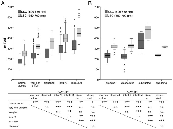
Figure 5.
Boxplot showing the prolongation of mean autofluorescence lifetimes (τm) for ectopic RPE. (A) RPE cells with uniform layer morphology (normal aging), very nonuniform RPE cell layer, sloughed RPE released into the subretinal space, and ectopic RPE intraretinal in the photoreceptor layer (intraPS) or after crossing the external limiting membrane (intraELM). (B) Bilaminar RPE, dissociated RPE in atrophic areas, subducted RPE cells and shedding RPE. The excitation wavelength was 960 nm (TPE), statistic by Kruskal-Wallis with Dunn-Bonferroni post hoc test. ***P < 0.001; **P < 0.01; *P < 0.05.

