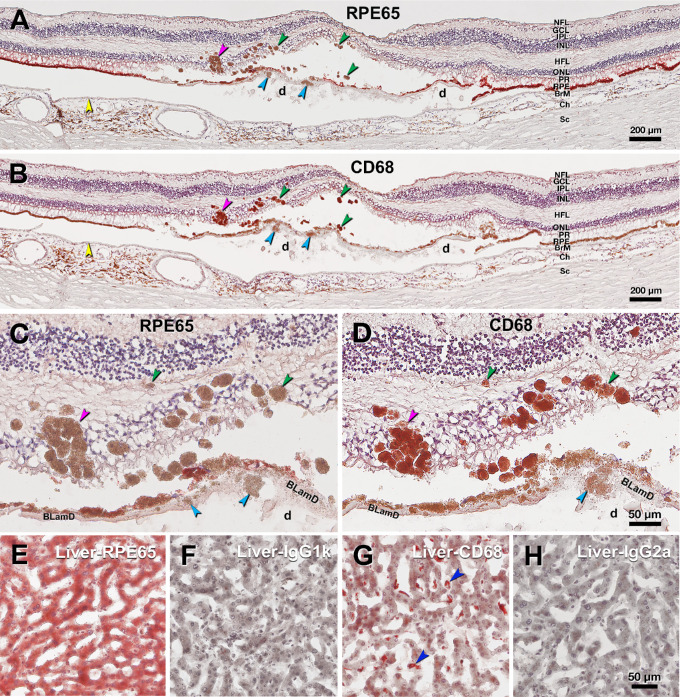
Figure 6.
Immunohistochemistry contrasts ectopic and orthotopic RPE in age-related macular degeneration. This retina of a 90-year-old white female donor has huge soft drusen and continuous basal laminar deposit (BLamD) under the fovea. Yellow arrowheads, Bruch's membrane. Scale bars apply to each row. (A) RPE65 immunoreactivity of ectopic RPE including sloughed/intraretinal phenotypes presenting as hyper-reflective foci (HRF) at green/pink (RPE plume) arrowheads and shedding RPE phenotype at cyan arrowhead. (B) CD68 immunoreactivity of ectopic RPE including sloughed/intraretinal phenotypes at green/pink (RPE plume) arrowheads and shedding RPE phenotype at cyan arrowhead in the same retina in panel A. (C) Magnified view of ectopic RPE lacking RPE65 immunoreactivity in panel A. (D) Magnified view of CD68 immunoreactive ectopic RPE in panel B. (E-G) Human liver tissue. E RPE65 positive control. F Sham control for RPE65. G CD68 positive control, blue arrowheads indicate CD68+ Kupffer cells. (H) Sham control for CD68. d, soft drusen; BLamD, basal laminar deposit; NFL, nerve fiber layer; GCL, ganglion cell layer; IPL, inner plexiform layer; INL, inner unclear layer; HFL, Henle fiber layer; ONL, outer nuclear layer; PR, photoreceptor layer; RPE, retinal pigment epithelium; BrM, Bruch's membrane; Ch, choroid; Sc, sclera.

