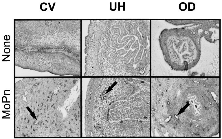
FIG. 2.
Expression of VCAM-1 in different regions of the GT following MoPn infection. Frozen sections of GTs prepared from uninfected (top panels) and infected mice (bottom panels) were stained with an anti-VCAM-1 monoclonal antibody and visualized using immunoperoxidase histochemistry. Venules staining positive for VCAM-1 can be seen in the CV region 7 days after infection (arrow in bottom left panel), in the UH on day 14 (arrow in bottom middle panel), and in the OD by day 21 (arrow in bottom right panel).

