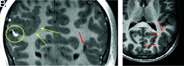
FIG 4.

An 18-year-old man with HHT. Coronal 3D T1 postcontrast imaging shows right parietal polymicrogyria (A, yellow arrows), an AVM (A, yellow circle), and a small AVM (A, red arrow). Axial T1 image shows left parietal occipital polymicrogyria (B, red arrows).
