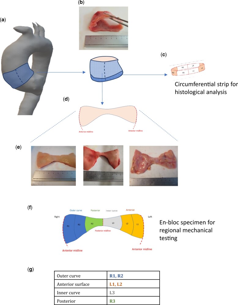
Figure 1:
Origin of the ascending thoracic aortic aneurysm specimen and estimated anatomical regions. The proximal ascending thoracic aortic aneurysm specimen is excised en-bloc (A and B). From the inferior-most border, a 4–5-mm strip of tissue is removed and processed for histological analysis ©. The remainder of the specimen is divided vertically down the anterior midline giving the characteristic butterfly appearance (D). Photographs of actual patient specimens are shown in E (intimal surface facing up). The circumferential regions for subsequent analysis are 6 equal regions (F), corresponding to the anatomical locations described in the table (G).

