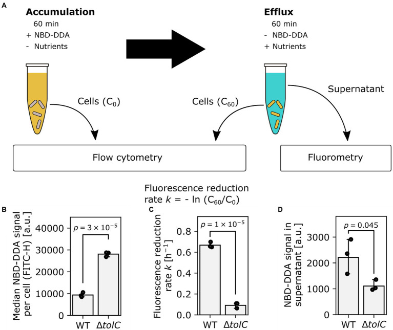
Figure 7.
TolC is involved in efflux of NBD-DDA. (A) Workflow to determine efflux activity in bacteria, using NBD-DDA. (B) Increased median accumulation of NBD-DDA per cell in E. coli ΔtolC as determined by flow cytometry suggests decreased efflux activity. (C) Reduced fluorescence reduction rate per cell and (D) NBD-DDA signal in the supernatant after 60 min are lower in E. coli ΔtolC, due to decreased efflux activity. p-values were obtained from unpaired, two-tailed t-tests of the fluorescence values or rates. Bars indicate mean of n = 3 replicates. Black dots indicate individual experiments. Errors are 95% confidence intervals of the mean obtained by bootstrapping.

