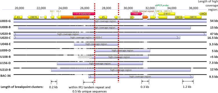
Fig 3. Overlap in KSHV IR1 overrepresentation regions.
Sequence alignment of regions with excess read coverage near IR1 (showing those with identical breakpoints only once). Location and orientations of ORFs are in yellow, long non-coding RNAs in red, and repetitive sequences in orange. Gene region used for ddPCR probe for screening is in green. The ’minor internal repeat’ is composed of the 13-bp sequence TGGGATGGGGGTG repeated 4 to 13 times. Vertical dashed red lines demarcate the minimal high coverage region. Indicated below are the sizes of the 4 breakpoint clusters identified. Breakpoint coordinates of the high coverage regions shown are indicated in S2 Table.

