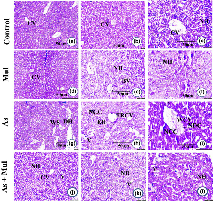
FIGURE 4.

Histological photograph of hepatic tissue of experimental mice (H & E stain). Hepatic tissues of control mice (a, b and c) showed normal hepatocyte (NH) with the central vein (CV). Mul‐treated mice liver (d, e and f) also showed normal hepatocyte (NH) with the central vein (CV) and blood vessels (BV). Hepatic tissue section of As‐treated mice (g, h and i) showed widened central vein (WCV), endothelial rupture in the central vein (ERCV), widened sinusoidal space (WS), enlarged hepatocyte (EH), nuclear degenerative changes (NDC), the appearance of severe necrosis in the cytoplasm (NCC), appearance in vacuoles (V) endothelial rupture in the central vein (ERCV), and degenerated hepatocytes (DH). Mul coadministration with As (As+Mul) (j, k and l) showed moderate changes with the central vein (CV), normal hepatocyte (NH), few appearances in vacuoles (V), and nuclear degeneration (ND). Microphotography of a, d, g and j in ×10; b, e, h, and k in ×20; and others in ×40 with hematoxylin and eosin stain
