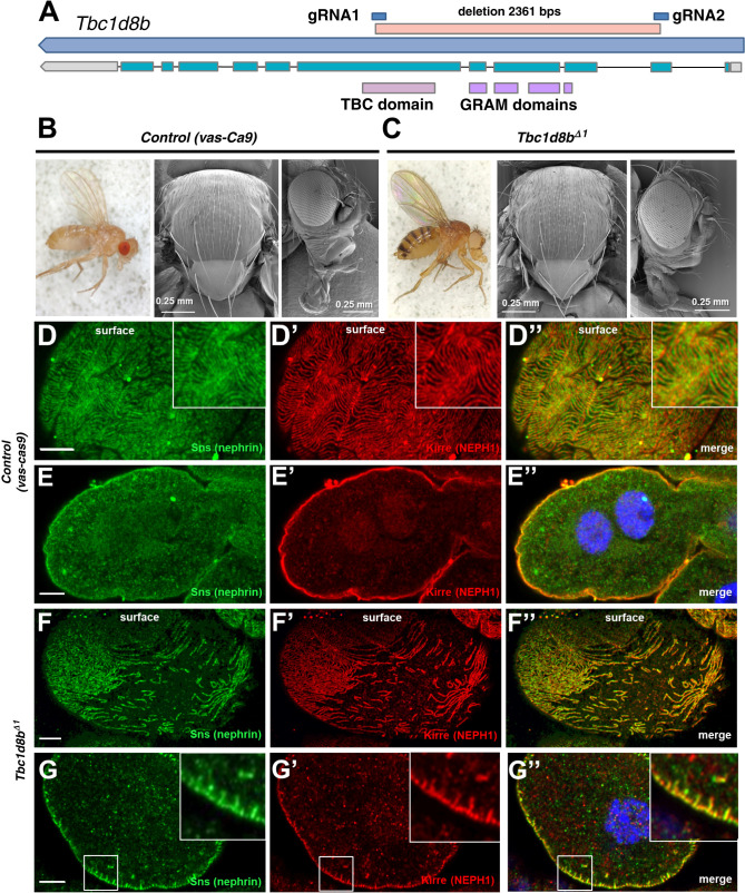
Figure 1.
A stable genetic deletion of Tbc1d8b in Drosophila shows a nephrocyte-restricted phenotype. (A) Schematic shows the Drosophila Tbc1d8b locus (CG7324) with the longest transcript and the protein’s functional domains corresponding to the respective exons. The deletion of the Tbc1d8bΔ1 allele is denoted between the respective gRNA target sites. (B) and (C) Stereomicroscopy of the whole fly (left) and scanning EM of thorax (middle) and head (right) are shown for control animals (vas-Cas9, B) and adult Tbc1d8bΔ1 allele flies (C). Tbc1d8b null animals show no overt phenotypic difference. (D)–(G″) Confocal microscopy of nephrocytes stained for nephrin (Sns) and Kirre (NEPH1) in tangential (D)–(D″) and (F)–(F″) or cross sections (E)–(E″) and (G)–(G″) are shown. Control cells (vas-Cas9, D–E″) show a regular slit diaphragm staining pattern, whereas Tbc1d8bΔ1 nephrocytes reveal a localized loss of slit diaphragm on the surface (F)–(F″) and small protrusions of slit diaphragm protein toward the interior of the cell (insets in G–G″). Nuclei are marked by Hoechst 33342 in blue. EM, electron microscopy.

