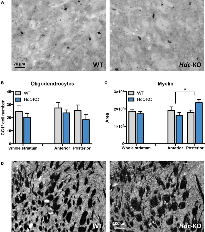
FIGURE 3.
Oligodendroglia and myelin in histidine decarboxylase-knockout (Hdc-KO) mice. (A) Representative images of CC1 immunostaining of the striatum in wild-type and Hdc-KO mice. (B) There was no significant difference between KO and WT mice in the number of mature oligodendrocytes. N = 5 WT, 5 KO. Two-way ANOVA: main effect of region, F(1,17) = 0.92, p > 0.35; main effect of genotype, F(1,17) = 2.1, p > 0.16; and interaction, F(1,17) = 0.18, p > 0.6. (C) White matter cross-sectional area, evaluated by Myelin Basic Protein (MBP) immunostaining, was elevated specifically in the central striatum. N = 6 WT, 6 KO. Two-way ANOVA: main effect of region, F(1,25) = 0.53, p > 0.45; main effect of genotype, F(1,25) = 2.3, p = 0.15; interaction: F(1, 25) = 4.630; p = 0.04. (D) Representative micrographs of WT and KO striata immunostained for myelin. All data are presented as mean ± SEM. *p < 0.05 by Sidak’s post-hoc test.

