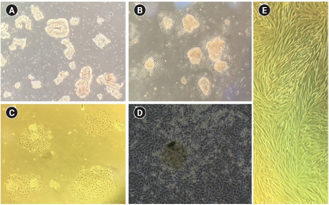
Figure 2.

Light microscopy of endometrial cells. (A) Epithelial glands. (B) Initial attachment of epithelial cells into the flask. (C) Island-shaped clusters of epithelial cells. (D) Confluent monolayer of epithelial cells with a dome-like structure. (E) Confluent stromal cells. ×50 magnification.
