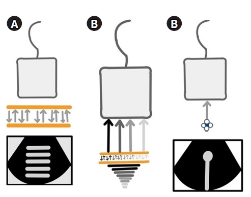
Figure 1.

(A) Reverberation artifacts [2]. When two reflective parallel interfaces lie in the path of the ultrasound beam, the beam gets reflected multiple times between these interfaces. Hence, the transducer assumes them to arise at increasing depths depending on the time delay between transmitted pulse and received echo. This generates an image comprising of multiple echogenic parallel lines placed equidistant from each other, which decrease in brightness with increasing depth. In the chest, the pleural line lies parallel to the probe and acts as a reflective interface when the lungs are aerated, giving rise to reverberation artifacts known as “A-lines” due to repetitive reflections between it and the transducer. Hence, the presence of A-lines denotes an aerated lung. It is important to note that tissue harmonic imaging (THI) will decrease this artifact. (B) Comet-tail artifacts [3]. These are a subtype of reverberation artifacts arising from highly reflective, closely apposed interfaces like calcium, cholesterol crystals, and metal, which cannot be resolved separately. The comet-tail artifact appears as a tapering echogenic cone, showing decreased amplitude and width with increasing depths. Previously, the B-lines were called comet-tail artifacts. (C) Ring-down artifacts [2]. These are similar to comet-tail artifacts but arise from fluid trapped between air. B-lines have been described to be ring-down artifacts. Comet-tail and ring-down artifacts are minimized by spatial compound imaging, which needs to be turned off to get less divergent, sharper, and brighter B-lines. THI also assists in producing sharper comet tails. Permission obtained from RSNA to reproduce images in print or web.
