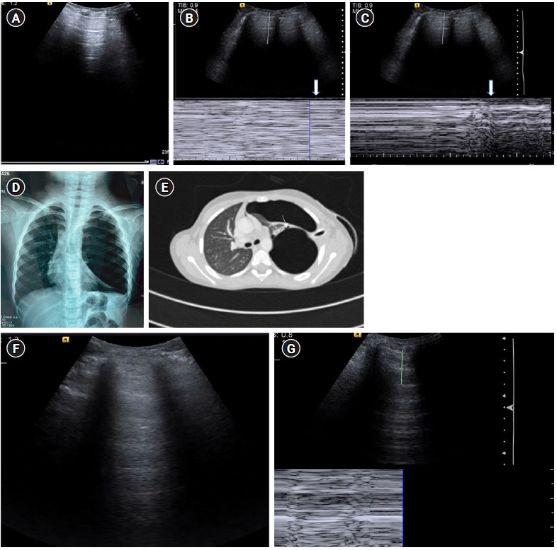
Figure 11.

(A-E) A’ profile. Sagittal grey scale ultrasound scan (A) with M-mode tracing (B) in a 6-year old boy with history of dyspnea, chest pain and previous intercostal chest tube drainage for empyema revealed abolished lung sliding in the form of “stratosphere sign”: horizontal lines above and below the pleural line, in the left anterior and lateral lung zones with presence of A-lines and absent B-lines (known as A’ profile). (C) Diagnosis of pneumothorax was confirmed on ultrasound by localizing the “lung point”: visualizing intermittent stratosphere sign and seashore sign (arrows). The lung point was however visualized in posterolateral lung zones, indicating a large pneumothorax. (D) Chest X-ray in the same patient revealed a large left sided pneumothorax with contralateral tracheomediastinal shift, further confirming the findings of a large pneumothorax as diagnosed on ultrasound. The patient was however hemodynamically stable. (E) Contrast-enhanced chest computed tomography scan chest revealed a bronchopleural fistula (arrow). (F) Cine US clip showing absent lung sliding (Supplementary Material 4). (G) Cine US clip showing “stratosphere sign (Supplementary Material 5).”
