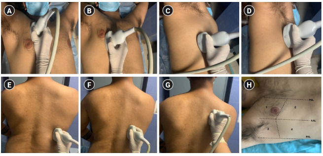
Figure 2.

Focused transthoracic ultrasound using 14-zone scanning protocol by Laursen et al. [6], based on principles of Lichtenstein and Volpicelli, using a curved array low-frequency transducer placed in a longitudinal axis over an intercostal space with the patient in a supine position. (A) Zone R1: transducer placed in the 2nd intercostal space, a few centimeters away from the sternum. (B) Zone R2: transducer placed in the 4th intercostal space in the mid-clavicular line. (C) Zone R3: transducer placed in the mid-axillary line on the lower part of the lateral chest, in approximately the 5th or 6th intercostal space. (D) Zone R4: transducer placed in the mid-axillary line on the upper part of the lateral chest, in the 3rd intercostal space. (E) Zone R5: with the patient in a sitting position, the posterior chest wall is scanned by placing the transducer on the lower part of the chest along a vertical line passing through the inferior angle of the scapula. (F) Zone R6: the transducer is placed medially in the posterior midclavicular line at a level corresponding to middle to lower part of the scapula. (G) Zone R7: transducer placed further cranially, medial to the upper part of the scapula. Similarly, scanning is done on the left side in zones L5–L7. (H) Zone 8 scanning of anterolateral chest wall proposed by Volpicelli et al. Zones are divided into anterior and lateral by the parasternal line (PSL), anterior axillary line (AAL), posterior axillary line (PAL), and upper and basal by a line passing below/at the level of the nipple-areola complex. Zone 1: anterior upper; zone 2: anterior basal; zone 3: lateral upper; zone 4: lateral basal.
