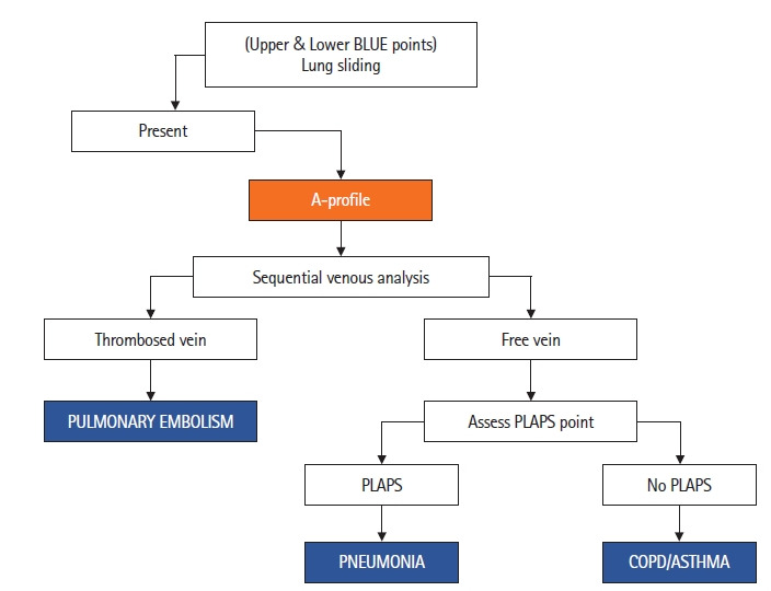
Figure 6.

The presence of lung sliding and A-lines constitutes the A-profile [7]. This is followed by sequential venous analysis to look for thrombosed veins (pulmonary embolism) or assessment of posterolateral alveolar and/or pleural syndrome (PLAPS) point in case of free veins to diagnose pneumonia (PLAPS) and chronic obstructive pulmonary disease (COPD)/asthma (no PLAPS). BLUE: bedside lung ultrasound in Emergency.
