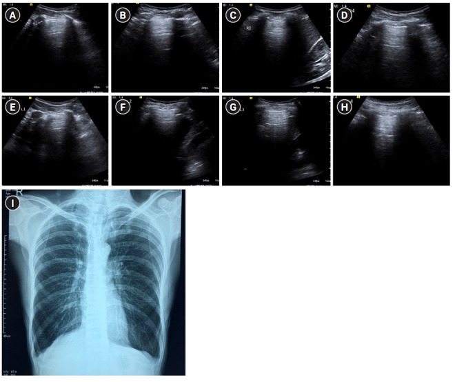
Figure 7.

A profile. Sagittal greyscale ultrasound scans (A-H) of eight lung zones in bilateral anterolateral chest wall revealing the A-profile A-lines with lung sliding and absence of B-lines in a 30-year-old afebrile male with a history of chronic cigarette smoking, presenting with dyspnea, diagnosed as exacerbation of chronic obstructive pulmonary disease/asthma on ultrasound. (I) Findings on chest X-ray were seen to be concordant. Bilateral lung fields appear hyperinflated with rounded costophrenic angles and flattening of hemidiaphragm.
