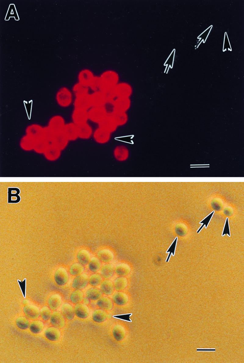
FIG. 1.

The C3.1 epitope is uniformly distributed on the cell surface of yeast forms of C. albicans. Hydrophilic stationary-phase yeast cells were reacted with MAb C3.1, washed, and counterreacted with fluorescence-labeled anti-mouse IgG. The C3.1 epitope is located over the entire cell surface (A). The same field was photographed under phase-contrast microscopy (B). The C3.1 epitope distribution displays the same homogeneous pattern over the cell surface as the epitope recognized by the IgM protective antibody, MAb IgM B6.1. These results provide further evidence that the IgG3 antibody is specific for the same oligomannoside as the protective IgM. Note that some yeast cells (arrows) and all of the blastoconidia (arrowheads) were nonreactive with MAb C3.1. The importance of nonreactive cells in pathogenesis and protection experiments is unknown. Bar, 10 μM.
