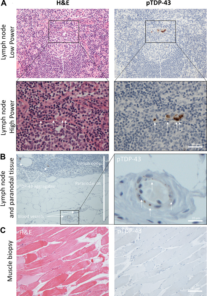
Figure 4.

pTDP‐43 aggregates identified in lymph node parenchyma, endothelial cells, and chondrocytes long before symptom onset of ALS. (A) H&E (left) and pTDP‐43 (right), low power (top) and high power (bottom) images of an active lymph node germinal centre with evidence of cells containing pTDP‐43 aggregates (white arrows) within the mantle zone rim of the lymphoid follicle. (B) Low power (left) and high power (right) images of the paranodal tissue demonstrating blood vessel pTDP‐43 aggregation (white arrows) in adjacent feeder vessels. (C) H&E (left) and pTDP‐43 (right) images showing no evidence of pTDP‐43 aggregates in a muscle biopsy taken at point of diagnosis from patient 1.
